
Shunyata Research in Electrophysiology: A Breakthrough in Measured Noise Reduction
Shunyata Research in electrophysiology
Electrophysiology is a term used to describe complex, catheterized heart surgeries. Cardiac surgeons use this procedure to treat and correct abnormal heart rhythms called arrhythmias. Surgeons performing this delicate procedure rely on highly resolved heart signals to successfully treat patients. Unfortunately, noise interference has wreaked havoc on the resolution of heart signals since electrophysiology procedures began in the mid 1980’s.
Signal resolution, a unifying goal
A common objective in electrophysiology, professional recording and sound reproduction is the critical resolution of low- amplitude signals. In each context, exposure to proximally generated noise will degrade signal resolution. Shunyata Research’s unique science possesses a measurably superior ability to mitigate high-intensity system generated noise, thus preserving the low level signal. The clinical measurements below show in visual terms what award-winning studios, recording engineers and record producers have noted for over 20 years. Whether you believe your eyes or your ears, it is clear that Shunyata Research designed power systems deliver superior results.
The Discovery
In 2015, celebrated electrophysiologist Dr. Daniel Melby, Lab Director at Minneapolis Heart Hospital, made a startling discovery when he tested Shunyata Research products in his lab. Dr. Melby documented his results and later went on camera to explain his findings when using a Shunyata-designed noise reduction system. Soon, a new company was formed — and the products evolved into what they are today. In 2016, Shunyata Research created a sister company named Clear Image Scientific®, which designs and manufactures meticulously-engineered power distribution systems for medical and science-based applications. CIS® power cords and power distributors have since been proven to measurably lower the multiple layers of noise that can affect ultra-sensitive, low- level signal acquisition systems.
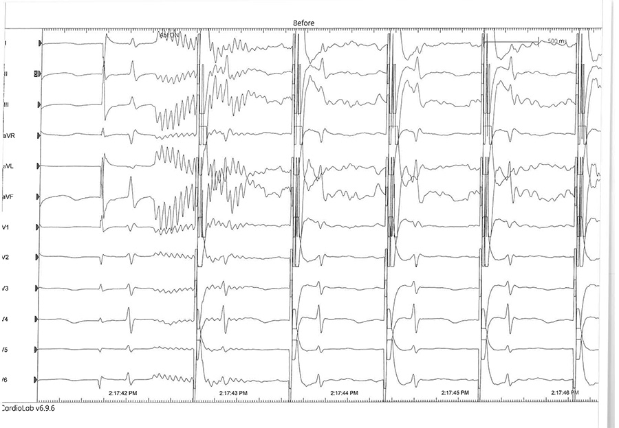
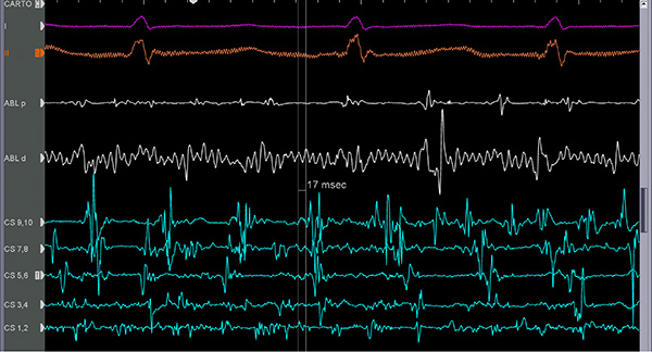
COMPARISON BEFORE: Screen 1
Depicts heart activity with unaddressed electrical noise on the line with electronics connected to an isolation transformer.
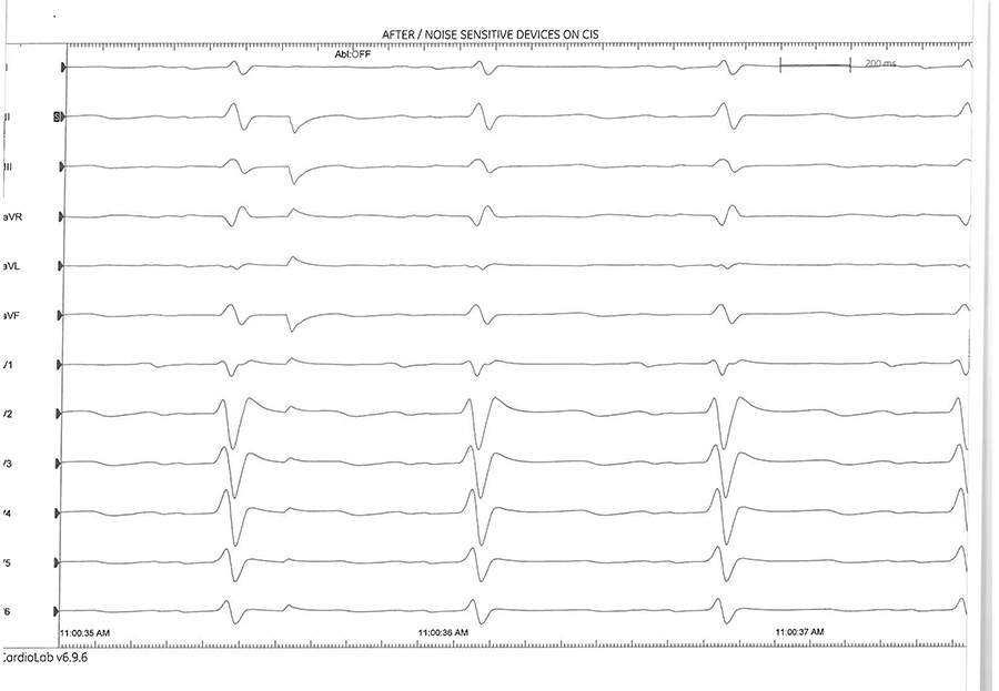
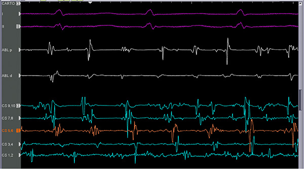
COMPARISON AFTER: Screen 2
Depicts the same conditions, but with monitoring equipment and recording amplifiers plugged into Clear Image Scientific® power distribution and shielded power cords.
Following is a small sampling of results from electrophysiology labs.
These measurements and experience based comments explain why Shunyata Research products have become the most technically sophisticated and scientifically-vetted products in their category.
Norton Health Hospital:
Louisville, KY
“After installing the CIS® units, the bipolar noise levels on the mapping catheters went from fairly high at .13 —.15 to less than 0.01 mV after installing the first of the two CIS® units and remained at that level after installing the second unit. As I mentioned before, 0.01 is the lowest the system can measure so the noise level for the bipolar signals is now below the threshold of what the system can measure.”
~ Dr. Kent Morris, Electrophysiology Lab Director: Norton Health Hospital, Louisville, KY
Measurable, consistent results with CIS® Minneapolis Heart Institute:
Minneapolis, MN
Noise: “In regards to noise, which I define as baseline artifact — the noise level on the mapping computer was the lowest I have seen in 10 years and over thousands of cases. In fact, the noise level was below my measurement capabilities.”
- Measured noise level before using CIS® products: 0.050 - 0.250mV
- Measured noise level after using CIS® products: dropped below measurement floor, estimated at 0.001 - 0.003mV
Resolution: “In regards to the equally important resolution of cardiac signals, which I define as observed voltage changes over time; on the mapping computer this also was the best that I have ever seen in my lab or any other lab in the world.”
Before: Resolution of mapping system was worse than the recording system.
After: The resolution with CIS® was better than the recording system by a wide margin.
- Signal amplification and processing used via CIS® power products outperformed our dedicated $75K signal amplifier.
- We recorded dramatic reductions in noise-levels as more CIS® products were applied.
- Best case of the day was the final procedure with the maximum number of CIS® products applied.
“The CIS® power conditioners and cords are now a permanent part of the operating room systems at the Minneapolis Heart Institute and have been recommended to countless other hospitals based on their astounding success.”
~ Dr. Daniel Melby, Medical Director, Electrophysiology Labs: Minneapolis Heart Institute, Minneapolis, MN
Scripps Cardiovascular Institute:
La Jolla, CA
“The results were impressive and consistent. This lab is now better than our recently built state-of-the-art labs. During our most complex ablations we are able to maximize gain without being troubled by noise. We are able to widen our filter settings. We are able to clearly see signal degradation during ablation on a more consistent basis than ever before.”
~ Douglas Gibson MD, FHRS, Director, Cardiac Electrophysiology: Scripps Clinic and Prebys Cardiovascular Institute, La Jolla, CA
Deborah Heart and Lung Center:
Browns Mills, NJ
“All of our EP Staff and surgeons have noticed an improvement in the signal quality in each of three labs after the application of CIS® systems. I believe we have finally found a product that can help eliminate noisy signals in our EPS Labs At Deborah Heart and Lung Institute.”
~ Garry Chamberlain, REMS, Assistant Director, Biomedical Engineering: Deborah Heart and Lung Center, Browns Mills, NJ
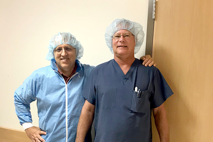
Jefferson Health: Philadelphia, PA
“After installing the CIS® system, we now have the cleanest 64 unipolar signals that I have seen in any of the labs that are doing Topera Rotor based Mapping. Clean signals are critical for all of the Biosense algorithms to work well. You need clean unipolar signals to accurately find the local timing on the bipolar signal. Further clean signals make sure ripple mapping is showing you true fractionated signals not noise. We’ve never seen signals this clean before.”
~ Dr. Bradley Bacik, DO FACC, FHRS, Director of Electrophysiology: Jefferson Health, Philadelphia, PA
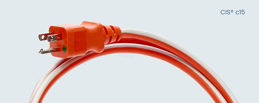
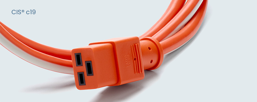
Cox South Hospital: Springfield, MO
“After installing the CIS® system and during ablations, we noted the lowest measurable noise in our labs. We were able to perform a procedure on the next patient without the use of an IVC electrode. The change and lowering of noise during cases is obvious.”
~ Michael Gleason RT(R)(CI)(VI) RCIS CEPS Clinical Account Specialist KC POD Great Lakes Region: Cox South Hospital, Springfiled, MO
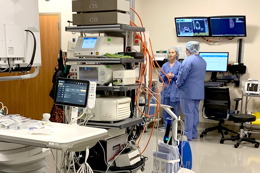
Einstein Medical Center: Philadelphia, PA
“We have been thrilled at the impact that CIS® had on the quality of our signals in our first lab. The system was easy to install and is unobtrusive. The company service is outstanding. As a result of the favorable impact, we requested installation in our second lab and expect to see similar results. ”
~ Sumeet K. Mainigi, MD, FHRS, Interim Chairman of Cardiology, Director of Electrophysiology: Einstein Medical Center, Philadelphia, PA
Documented CIS® results submitted to HRS Medical Journal
The following is a submission to the Heart Rhythm Society Journal made by a team of surgeons and engineers at Deborah Heart And Lung Center in New Jersey.
These A/B results were in direct comparison to standard transformer based power conditioning systems — during an active surgical procedure.
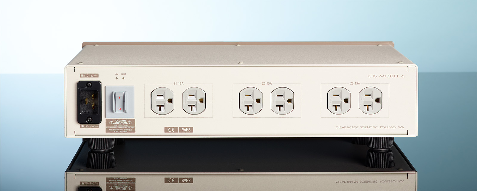
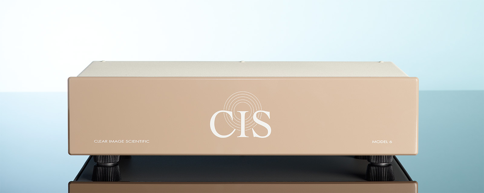
This document was submitted to the Heart Rhythm Society Journal, in which the lab team characterized the CIS® Model 6 as a “significant new discovery” in electrophysiology.
(Clear Image Scientific® is a sister company created by Shunyata Research to serve the medical and scientific communities.)
Independent noise test with CIS® Model 6
Signal noise reduction on electrophysiology amplifier utilizing Clear Image Scientific® Model 6
Test Conducted at Deborah Heart and Lung Center: Browns Mills, NJ
Participants: Pedram Kazemian, MD FACC; Raffaele Corbisiero, MD FACC; George Giacono, RN; Gary Chamberlain, REMS; David Muller, RN
Background: Noise on intracardiac recording systems has been a consistent problem since the inception of Electrophysiology Labs. External environmental sources and equipment is often a source of noise due to RFI and EMI emission. Isolating equipment is difficult due to the wide variety of equipment used during a range of different procedures.
Objective:
We compared signal quality with and without the use of the Clear Image Scientific® Model 6 to compare noise in a real-world setting (Clear Image Scientific® Inc. is located Poulsbo, WA).
Methods:
After witnessing a consistent reduction in noise when using the CIS® Model 6 over a period of time, we recorded a patient undergoing an RF ablation with catheters in place. The signal noise amplitude was recorded on both surface and intracardiac electrogram’s (EGM) with the CIS® Model 6 in place. With catheters in still in place, the amplifier was rebooted after the CIS® M6 was removed and the measurements were repeated with the amplifier plugged into a common transformer outlet.
Results:
With the CIS® Model 6 in place, surface ECG baseline noise amplitude was measured at 0.009 with the gain/ filter Low/High/Notch set to 128/1Hz/25Hz/60Hz respectively and for the EGMs 0.010 with the gain/filter Low/High/ Notch set to 128/30Hz/250Hz/60Hz respectively. Upon repeating without the CIS® Model 6, ECG baseline noise amplitude was measured at 0.022 with the gain/filter Low/High/Notch set to 128/1Hz/25Hz/60Hz respectively and for the EGMs 0.016 with the gain/filter Low/High/Notch set to 128/30Hz/250Hz/60Hz respectively.
Conclusion:
The CIS® Model 6 reduced baseline noise by 144% on the surface and 37% on the EGM.
reduced baseline noise on the surface
reduced baseline noise on the EGM
Limited Warranty
Clear Image Scientific® warrants products to be free of manufacturing defects in materials and workmanship for a period of two-years from date of purchase, in accordance with exclusions and conditions.